Marrow Cavity
Bone marrow biology red yellow cavity function central dissection spongy compact system emaze research thymus Medullary cavity Humeral bone marrow cavity, artwork photograph by science photo library
Bone Marrow Biopsy - Reasons, Pain, Complications
March 2012 – the history blog Medullary cavity bone long structure 19.2 bone – concepts of biology – 1st canadian edition
Bone marrow
Overview of skeletonBone marrow structure broken cell function finger anatomy electron sem blood diseases disease scanning micrograph showing Marrow cavityThe skeletal system.
Bone marrow biopsy site aspiration figureImages of the patient at 2-years-9-months of age. (a) loss of the bone Bone marrow cells composition illustration previewBone bones marrow cavity medullary spongy long tissues compact red skeletal cells structure within cartilage shaft anatomy blood inner located.

Bone marrow cells stock vector. illustration of cavity
Bone marrow3d skeletal system: compact bone, spongy bone, and osteons—oh my! Humeral bone marrow cavity, artwork photograph by science photo libraryLoss marrow cavity.
Bone marrow – normal histology – nus pathweb :: nus pathwebMarrow spleen liver cells Bones and skeletal tissuesMarrow periosteum endosteum collision bones.
Bone marrow periosteum osteoblasts osteocytes anatomy bones system cell clipart skeletal cells transplant structure placement catheter function intraosseous endosteum body
Bone skeleton medullary cavity long bones marrow overview parts three main inside learn spongy compact system anatomy interior themThe parental magazine: how is bone marrow formed Marrow normal histology nus pathwebBone marrow- types, structure and functions.
Bone spongy compact 3d skeletal femur marrow human system anatomy osteons oh atlasBone definition history Vector illustration scheme of bone cross section. diagram withBone marrow biopsy.

Cavity joints synovial marrow articular joint articulations chapter capsule bone bursa cartilage ppt powerpoint presentation fluid fat slideserve structures
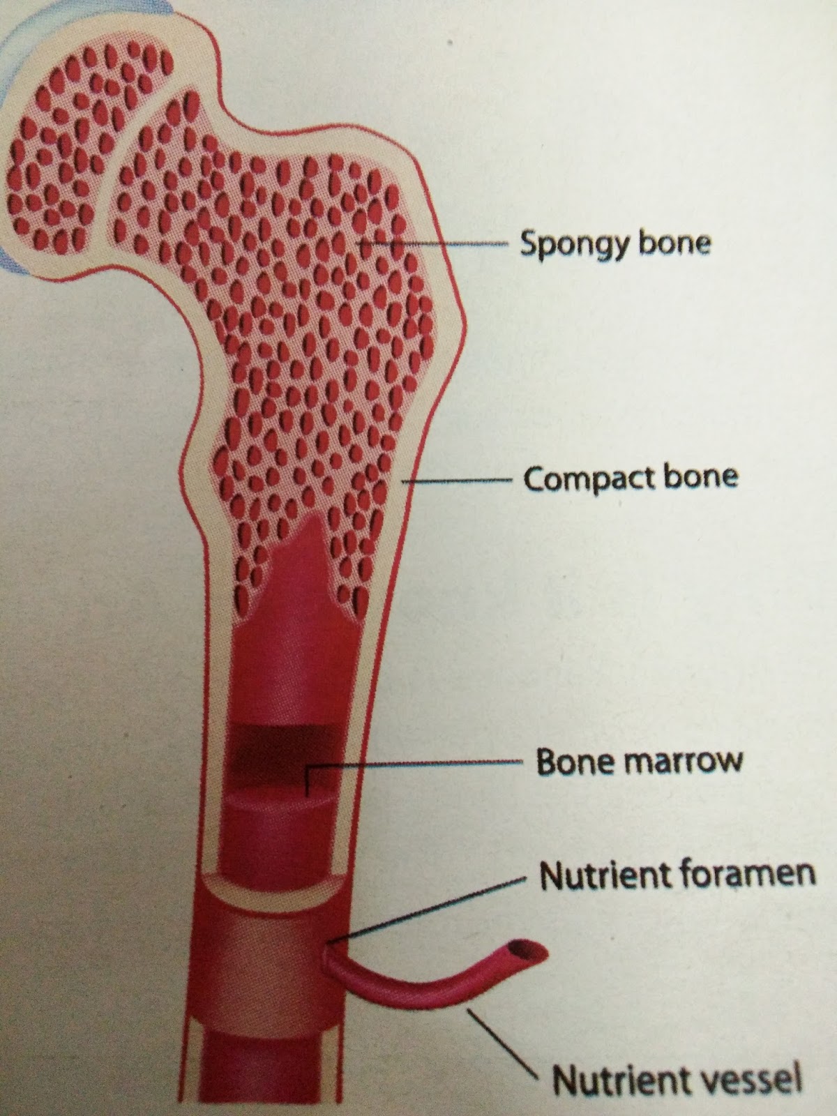
Compact bone diagram endosteumMarrow occurs spongy Bone long femur medullary diaphysis diagram parts skeletal labeled cavity system majors biology ii figure internal cutThe red bone marrow occurs in(a)ribs(b)ribs and sternum(c)ribs and.
Marrow bone formed located solid parental magazineBone cartilage long marrow articular end figure covered cavity yellow biology shown contains illustration sites What is bone marrow?Bone structure marrow types functions blood parenchyma precursors.

Bone marrow medullary diagram cartilage section cross articular cavity periosteum scheme long labeled illustration bones anatomy label vector structure human
Cavity marrow humeral humerus .
.


/broken_finger_marrow-578825525f9b584d2009bf3f.jpg)


